Mapping the Human Brain to Better Understand Neurological Diseases
Identifying and understanding structural changes in the brains of people affected by neurological conditions — such as Alzheimer’s disease, Parkinson’s disease, and multiple sclerosis, as well as those who have sustained a concussion — could lead to significant advances in patient care.
Through the Human Connectome project, Maxime Descoteaux’s research team aims to produce a complete map of the brain’s neural connections. This complex mapping would provide a deeper understanding of the functions of each brain region and become a valuable tool for research, clinical practice, and personalized medicine.
To achieve this, the team uses tractography, a diffusion magnetic resonance imaging (dMRI) technique. This method enables the study of the brain’s white matter — also known as the connectome — and allows for the reconstruction of a “virtual map” of the connections between different brain regions in a given individual. The researcher’s goal is to map the brains of 100,000 people.
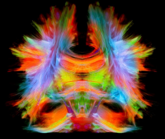
The task is no small feat: the human connectome contains approximately 160,000 km of nerve fibers!
The project goes even further, as the team aims to develop new AI-powered tractography tools to improve data quality and increase the speed of certain processing steps by a factor of 100.
“We are dealing with multidimensional, complex, and massive datasets, even at the scale of a single individual. Now imagine what that means for a database of 100,000 people! This requires tremendous computing power, including access to CPUs, GPUs, as well as large amounts of RAM and storage,” explains Maxime Descoteaux.
“A single dMRI scan is approximately 200 MB, tractography and processing pipelines require around 45 CPU-hours of computation, generate 15 GB of output data, and a complete connectome contains approximately 10 million connections (1 GB). The infrastructure provided by Calcul Québec and the Digital research Alliance of Canada is essential to carrying out a project of this scale,” he adds.
Ultimately, the Human Connectome project will be a major asset for clinicians working to diagnose their patients, enabling precise, reliable, and easily generalizable comparisons across populations.
Researcher Maxime Descoteaux is a member of the Medical Imaging axis at the CHUS Research Centre, a professor in the Department of Computer Science at the Faculty of Science of the Université de Sherbrooke, director of the Platform for Analysis and Visualization of Images (PAVI), and director of the SCIL laboratory (Sherbrooke Connectivity Imaging Lab).
This year, he was named a Senior Fellow of the International Society for Magnetic Resonance in Medicine (ISMRM). Photo credit : Université de Sherbrooke